Figure 4.
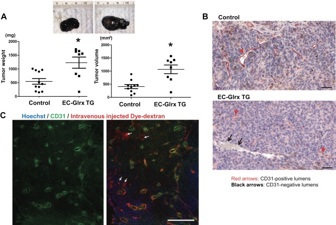
EC-specific Glrx overexpression promoted tumor growth and EC-independent tube formation in B16F0 tumors. A) B16F0 tumors from control and EC-Glrx TG mice, extracted after 2 wk of inoculation. Representative photos of tumors (top), and weight and volume assessments (n = 8–12). *P < 0.05. B) CD31 staining on the paraffin section of B16F0 tumors demonstrates EC-lining lumina (CD31-positive cells shown in red arrows) as well as blood cell–containing lumen without CD31 staining (shown by black arrows, mostly found in EC-Glrx TG tumors). Photos are representative sections of tumors from EC-Glrx TG and control mice (scale bars, 50 μm). C) Blood flow in a tumor without endothelium in Texas Red-labeled Dextran-injected EC-Glrx TG mouse. CD31 from tumor sections were stained in green (left). The merged photo (right) indicates dye-conjugated dextran in blood flow (red), CD31 in EC (green), and nuclear staining Hoechst 33342 (blue). White arrows point to Dextran-positive CD31-negative areas (scale bar, 500 μm).

