Figure 3.
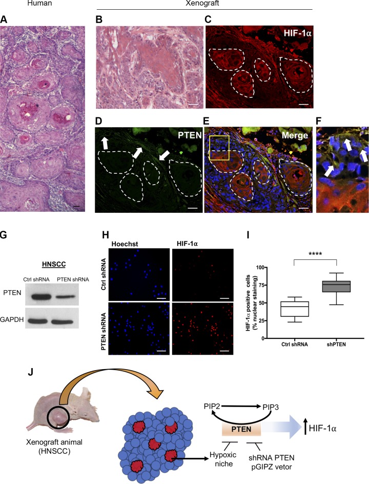
Genetic deregulation of PTEN induces HIF-1α expression. A) Tumor sample (H&E) from a human HNSCC depicts the formation of a typical morphologic aspect of a fast-growing HNSCC displaying the development of multiple tumor islands composed of concentric squamous malignant epithelial cells. B) Normal histologic architecture of xenograft-derived squamous cell carcinoma (H&E) presenting tumor islands depicting different degrees of cellular differentiation. C–F) Immunofluorescence staining of squamous cell carcinoma xenografts displaying HIF-1α staining conjugated with Alexa 568 (C), PTEN staining conjugated with Alexa 488 (D, arrows), merge of all 3 channels (E) presenting cells positive for PTEN within the tumor mass (F, arrows), and within the hypoxic niche, and hypoxic areas (red). G) Down-regulation of protein levels of PTEN upon delivery of PTEN-shRNA. H) Representative image of HNSCC cells expressing high levels of HIF-1α detected by immunofluorescence after delivery of PTEN-shRNA. I) Quantification of the total number of HIF-1α–positive cells from tumor cells receiving PTEN-shRNA compared with control shRNA (scrambled shRNA). ****P < 0.0001. J) Schematic representation of hypoxic niches and the expression of PTEN-driven expression of HIF-1α. Scale bars, 100 μm.

