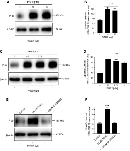
Figure 2.
PGH2 and PGE2 up-regulate P-gp protein and activity levels in isolated brain capillaries. A) Western blot showing P-gp protein expression in isolated rat brain capillaries exposed to PGH2; β-actin was used as protein loading control. B) P-gp transport activity in isolated rat brain capillaries exposed to PGH2. C) Western blot showing P-gp protein expression in isolated rat brain capillaries exposed to PGE2; β-actin was used as protein loading control. D) P-gp transport activity in isolated rat brain capillaries exposed to PGE2. E, F) Exposing brain capillaries to 25 nM PGH2 increased P-gp protein expression and transport activity levels. This effect on P-gp was blocked by 100 nM BI1029539. For specific luminal NBD-CSA fluorescence, data represent means ± sem for 10 capillaries from a single preparation (pooled tissue from 10 rats). Units are arbitrary units (au; scale: 0–255). ***P < 0.001, significantly higher than controls.

