Figure 6.
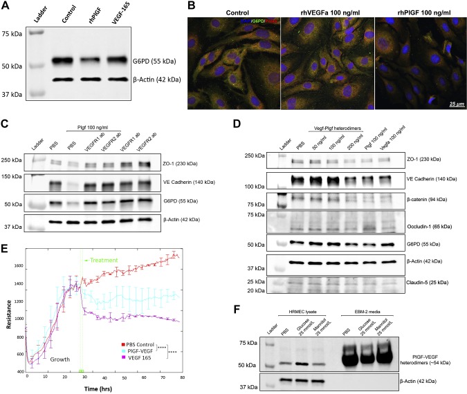
PlGF regulates G6PD, PRDX6, and EC barrier function through VEGFR1 and VEGFR2 signaling. A) WB results for G6PD. HRECs that were treated with rhPlGF and VEGF-165 protein (100 ng/ml each, for 2 d) were used for the WB assay: β-actin was used as the protein loading control. Note that rhPlGF, but not VEGF-165, down-regulated the protein level of G6PD compared with the PBS control. B) Double immunofluorescence labeling of G6PD and PRDX6. The HRECs that were treated with the PBS control, rhVEGF(a), and rhPlGF protein were fixed and stained with G6PD (green) and PRDX6 (red). The DAPI staining of the nucleus (blue) was used as the counterstaining. Note that the 2 proteins were colocalized in the PBS-treated and rhVEGF-treated cells (yellow); however, the staining signals were reduced in the rhPlGF-treated cells. The complete images of all 3 fluorescent staining channels are shown in Supplemental Fig. S5. C) WB results of ZO-1, VE-cadherin, and β-catenin. The confluent HRECs were treated with PBS, PBS + rhPlGF (100 ng/ml), rhPlGF + VEGFR1 antibodies (100 µg/ml), rhPlGF + VEGFR2 antibody (100 µg/ml), and VEGFR1 antibody alone or VEGFR2 antibody alone for 2 d. Note that rhPlGF protein down-regulated the protein levels of ZO-1, VE-cadherin, and G6PD, which were prevented by the VEGFR1 or VEGFR2 antibody. The antibody alone did not change the protein levels, which were equivalent to those of the PBS control. D) WB results of ZO-1, VE-cadherin, β-catenin, occludin-1, claudin-5, and G6PD. The confluent HRECs were treated with the PBS, PlGF/VEGF heterodimer (50, 100, or 200 ng/ml), PlGF (100 ng/ml), or VEGF-165 (100 ng/ml). Note that PlGF, the PlGF/VEGF heterodimer (200 ng/ml), and VEGF-165 down-regulated ZO-1, VE-cadherin, β-catenin, and claudin-5 (but not occludin-1); however, only PlGF and PlGF/VEGF down-regulated G6PD. E) TEER (or resistance) of HRECs. After the HRECs grew to confluence, the PBS control, PlGF/VEGF, or VEGF-165 was added to the medium, and the TEER was measured with ECIS in real time. The TEER values of the PlGF/VEGF heterodimer (blue) and VEGF-165 (purple) were significantly lower than those of the PBS control (red). Error bars represent sd out of 4 duplicate samples. F) WB results of PlGF/VEGF heterodimers in HREC lysate and culture medium. The confluent HRECs were treated with PBS, 25 mM d-glucose [high glucose (HG)], and 25 mM l-glucose [normal glucose (NG)]. Note that compared with the PBS controls, the HG up-regulated PlGF/VEGF in the cell lysates but decreased the levels in the culture medium. The β-actin was presented in the HREC lysate (but not in the culture medium) and used as the protein loading control. ****P < 0.0001.

