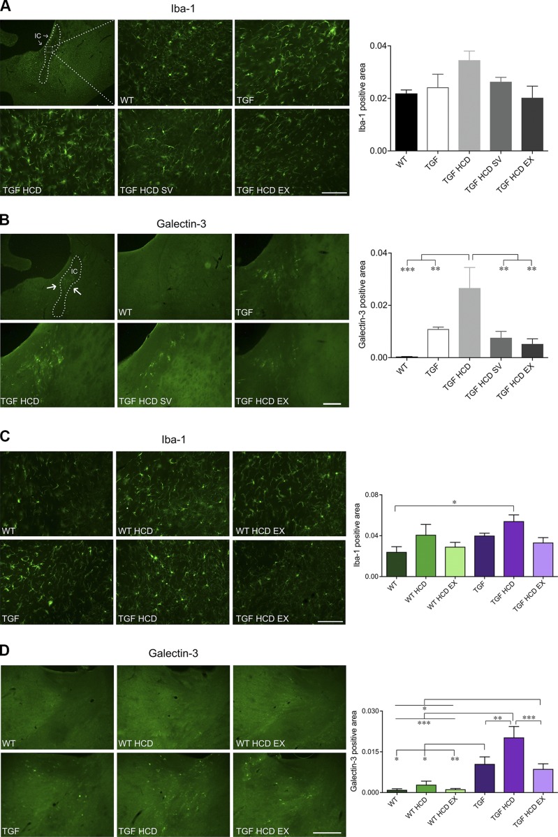
Figure 5.
A subset of microglia in WM was up-regulated by HCD and completely normalized by SV and EX. A, C) Iba-1–positive microglial area was quantified in the internal capsule (IC). In both cohorts, there was a tendency for an increase in microgliosis in HCD-fed mice and for SV and EX to prevent this response. B, D) In contrast, Gal-3–positive microglial area was selectively and significantly increased in TGF HCD mice in both cohort 1 (B) and cohort 2 (D). SV and EX, whether unlimited (B) or limited (D), fully countered this HCD-induced up-regulation of Gal-3 in TGF mice. Scale bars: 100 μm (Iba-1 images), 250 μm (Gal-3 images) (n = 4–5 mice/group). *P < 0.05, **P < 0.01, ***P < 0.001.

