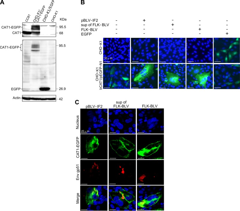
Figure 2.
CAT1/SLC7A1 protein acted as a cellular receptor and was required for BLV infection. A) Confirmation of the absence of endogenous CAT1 expression in CHO-K1 cells. CHO-K1 cells were transfected with and without bCAT1/pEGFP-N1 or with pEGFP-N1 as a negative control. The cell lysates were prepared 48 h post-transfection and then subjected to Western blot analysis using anti-CAT1 (upper panel), anti-EGFP (middle panel), and anti–β-actin (lower panel) antibodies. CC81 cell lysates were used as a positive control. The positions and MW of CAT1-EGFP, CAT1, EGFP, and actin are indicated. B) bCAT1/SLC7A1 expression associated with BLV cell-free and cell-to-cell infection. CHO-K1 cells were transfected with bCAT1/pEGFP-N1 expression plasmid with or without the BLV infectious molecular clone pBLV-IF2 or cocultured with supernatants from FLK-BLV cells or FLK-BLV cells at 4 h post-transfection. CHO-K1 cells were transfected with bCAT1/pEGFP-N1 or with pEGFP-N1 as a negative control. After 48 h incubation, the cells were fixed with 3.7% formaldehyde and stained with 10 µg/ml Hoechst 33342. EGFP-expressing syncytia were evaluated and quantitated using EVOS2 fluorescence microscopy. C) Detection of CAT1 and viral protein Env in syncytium. The transfected CHO-K1 cells were labeled with an anti-gp51 mAb (BLV-1) followed by incubation with Alexa Fluor 594 goat anti-mouse IgG (red). Nuclei were stained with Hoechst 33342 (blue). CAT1-EGFP was visualized to determine CAT1 expression (green) in the cells.

