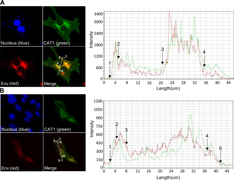
Figure 4.
Colocalization of CAT1/SLC7A1 and BLV Env protein in endomembrane compartments and at the cell membrane. bCAT1/SLC7A1 expression plasmid bCAT1/pEGFP-N1 was transfected into both FLK-BLV cells (A) and PK15/pBLV-IF2, which was stably transfected pBLV-IF2 (B); the transfected cells were grown on cover glasses and labeled with an anti-gp51 mAb (BLV-1) followed by incubation with Alexa Fluor 594 goat anti-mouse IgG (red). Nuclei were stained with Hoechst 33342 (blue). CAT1-EGFP was visualized to determine CAT1 expression (green) in the cells. The cells were evaluated, and the mean fluorescence intensities were visualized and analyzed along the line using an Olympus FV1000 laser-scanning confocal fluorescence microscope. White arrows and numbers indicate that positions of endomembrane compartments and the cell membrane.

