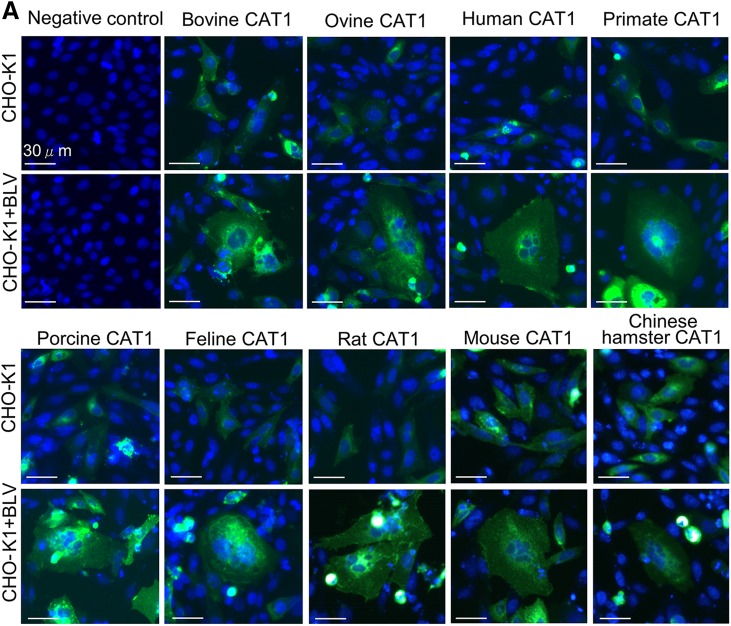
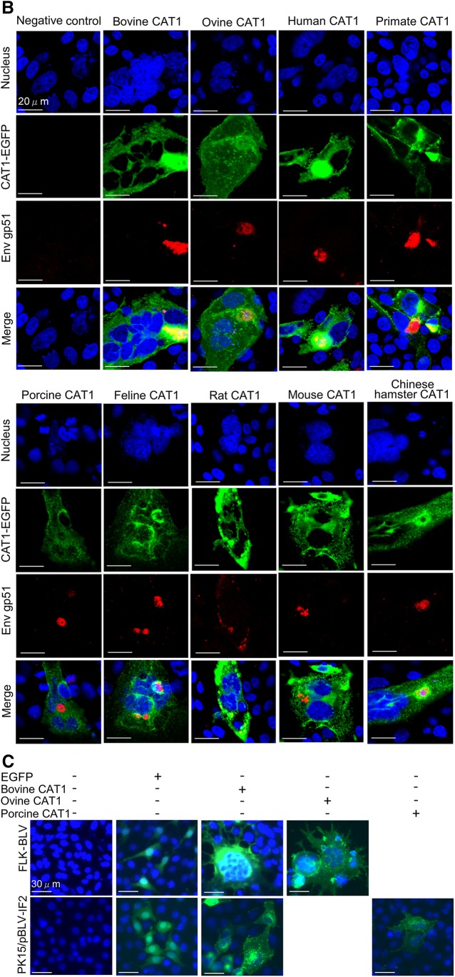
Figure 6.
CAT1/SLC7A1 did not have species specificity with all demonstrating susceptibility to BLV infection. A) CAT1/SLC7A1s from 9 different animal species did not have species specificity for BLV infection. The pEGFP-N1–expression plasmids encoding different CAT1/SLC7A1 proteins were derived using various animal species, including cattle (bovine), sheep (ovine), human, monkey (primate), pig (porcine), cat (feline), rat, mouse, and Chinese hamster. The plasmids were constructed and transfected into CHO-K1 cells. After 4 h transfection, the cells were added to culture supernatants of FLK-BLV cells and cultured for 48 h. The cells were then fixed with 3.7% formaldehyde and stained with 10 µg/ml Hoechst 33342. EGFP-expressing syncytia were detected using EVOS2 fluorescence microscopy. B) Detection of CAT1 and viral protein Env in syncytium. After 48 h incubation with the supernatant of FLK-BLV cells, the transfected CHO-K1 cells were labeled with an anti-gp51 mAb (BLV-1) followed by incubation with Alexa Fluor 594 goat anti-mouse IgG (red). Nuclei were stained with Hoechst 33342 (blue). CAT1-EGFP was visualized to determine CAT1 expression (green) in the cells. C) Ovine, porcine, and bCAT1/SLC7A1 proteins formed syncytia in both FLK-BLV and PK15/pBLV-IF2 cells following BLV infection. The FLK-BLV cells and PK15/pBLV-IF2 cells that stably transfected pBLV-IF2 were transfected with either ovine CAT1/pEGFP-N1, porcine CAT1/pEGFP-N1, bCAT1/pEGFP-N1, or pEGFP-N1 and incubated for 48 h. The cells were then fixed with 3.7% formaldehyde and stained using 10 µg/ml Hoechst 33342. EGFP-expressing syncytia were detected using EVOS2 fluorescence microscopy.


