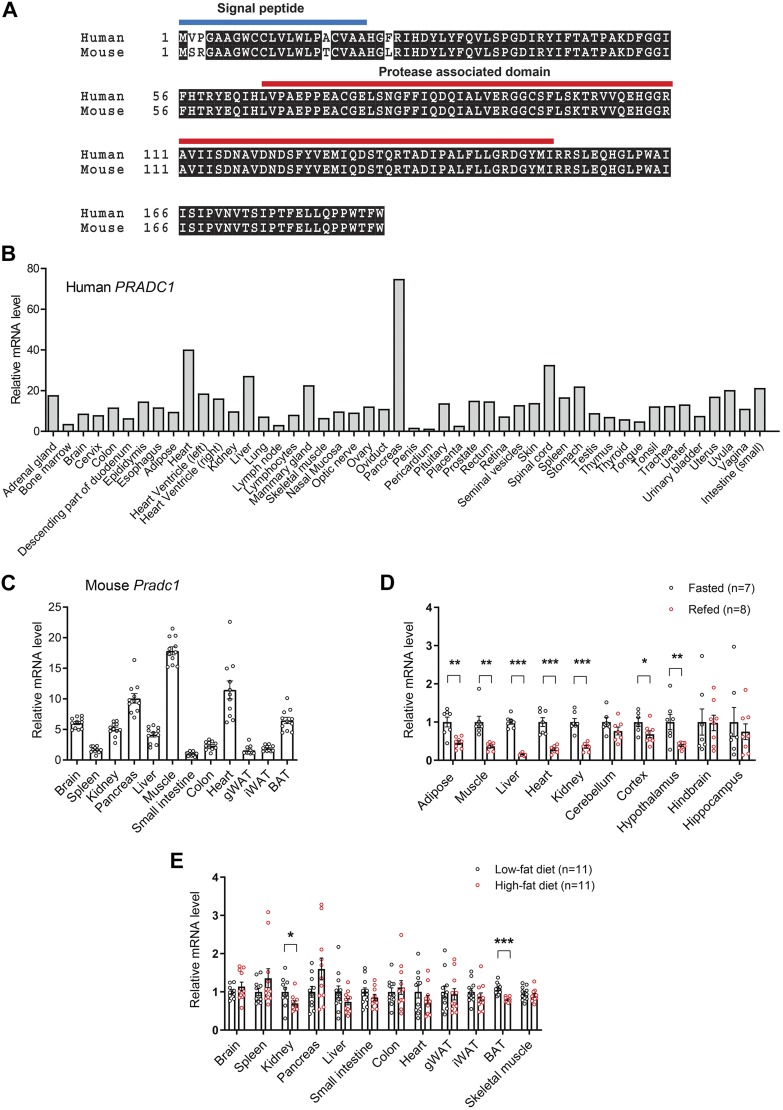
Figure 1.
Expression and regulation of Pradc1 in various tissues under different physiologic states. A) Sequence alignment of full-length human (NP_115695) and mouse (NP_001156899) PRADC1 proteins. The predicted signal peptide and protease-associated domain are indicated. B) Expression of PRADC1 in human tissue panel. C) Expression of Pradc1 in mouse tissues (n = 11). D) Real-time PCR analysis of the expression of Pradc1 in different tissues and brain regions from mice subjected to overnight food restriction (unfed group; n = 7) or overnight food restriction followed by 3 h of refeeding (refed group; n = 8). E) Real-time PCR analysis of the expression of Pradc1 in different tissues from mice fed a control LFD (n = 11) or an HFD (n = 11). Expression levels were normalized to β-actin. Data are expressed as means ± sem. *P < 0.05, **P < 0.01, ***P < 0.001.

