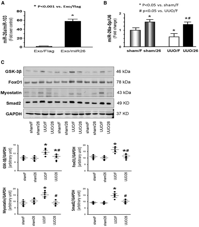
Figure 4.
Exogenous exosome–carried miR-26 suppressed UUO-induced muscle atrophy. A) Exosomes were harvested from conditioned medium from established HEK293–miR-26a or HEK293-Flag cell lines. Total RNA was isolated from these 2 groups. The expression of miR-26a-5p in exosomes was assayed by real-time qPCR. The bar graph shows expression levels of miR-26a in the HEK293–miR-26a group compared with the level in the HEK293-Flag group (designated as 1-fold). Results are normalized to U6. Results are reported as means ± se; n = 6/group. *P < 0.001 vs. HEK293-Flag. B) Total RNA was isolated from the TA muscle of mice given a sham intramuscular injection with exosome-carried Flag (sham/F), mice given a sham treatment with exosome-carried miR-26a (sham/26), UUO mice treated with exosome-carried Flag (UUO/F), and UUO mice treated with exosome-carried miR-26 (UUO/26). The expression of miR-26a-5p was assayed by real-time qPCR. The bar graph shows miR-26a expression from each group compared with levels in the sham/F group (represented at 1-fold). Results are normalized to U6. Results are reported as means ± se; n = 6/group. *P < 0.05 vs. sham/F; #P < 0.05 vs. UUO/F. C) The proteins were isolated from the TA muscle of sham/F, sham/26, UUO/F, and UUO/26 mice. The proteins GSK-3β, FoxO1, myostatin, and SMAD2 were measured by Western blotting in different groups of mice. Top: representative immunoblots. Bottom: densitometry analysis. The point graphs show the change of each protein band normalized to GAPDH. Results reported as means ± se; n = 6/group. *P < 0.05 vs. sham/F; #P < 0.05 vs. UUO/F.

