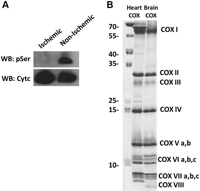
Figure 2.
Cytc purified from ischemic porcine brain tissue is dephosphorylated. A) Purified porcine brain Cytc from ischemic tissue (lane 1) and normal, nonischemic tissue (lane 2). Top, immunoblot with phosphoserine (pSer) antibodies; bottom, immunoblot with a Cytc antibody. B) COX purified from porcine heart (lane 1) and brain (lane 2) resolved on a high-resolution SDS-PAGE-urea gel showing all 13 tightly bound subunits of COX. WB, Western blot.

