Figure 3.
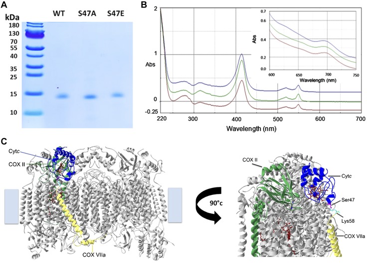
Characterization of recombinant WT, S47A, and S47E Cytc. A) Coomassie gel of bacterially overexpressed and purified recombinant WT (lane 1), S47A (lane 2), and S47E (lane 3) Cytc indicates that the proteins were purified to homogeneity. B) Reduced Cytc (5 µM) spectra (220–700 nm) and oxidized Cytc (100 µM) spectra (600–700 nm, inset) of recombinant WT (blue), S47A (red), and S47E (green) indicate correct folding and functionality of the proteins. C) Docking model of Cytc and COX (45, 60). A COX residue within a distance of 3 Å from residue 47 (magenta) is highlighted (Lys58 of COX subunit VIIa, cyan). Abs, absorbance.

