Figure 3.
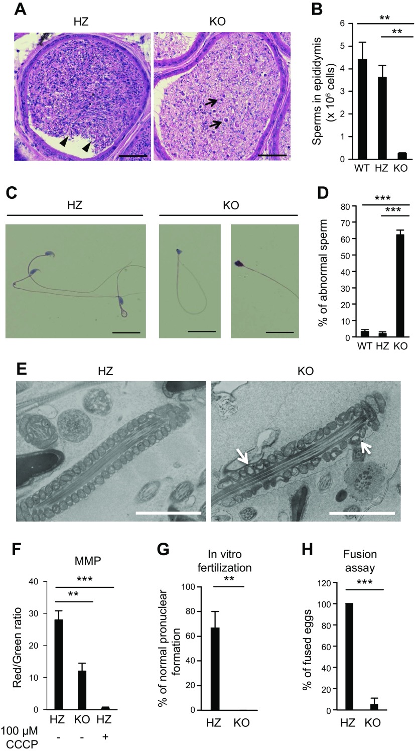
The function and quantity of ACSL6 KO sperm were disordered. A) Representative images of HE staining of the ACSL6 HZ and KO cauda epididymis. Arrowheads indicate normal sperm with tails and heads. Arrows point to cell debris for the KO mice. B) The numbers of WT, HZ, and KO sperm from the cauda epididymis. Data are presented as means ± sem (n = 4). C) HE staining of HZ and KO sperm. D) Percentage of abnormal sperm from WT, HZ, and KO mice. Data are presented as means ± sem (n = 4). E) FE-SEM images of ACSL6 HZ and KO epididymis. The arrows show the mitochondria devoid of content. F) MMP measured by flow cytometry. G, H) Successful IVF with (G) or without (H) the ZP. Scale bars, 2 µm. Data are presented as means ± sem (n = 3). **P < 0.01, ***P < 0.001 [Tukey’s honestly significant difference (HSD) test (B, D, F); unpaired Student’s t test (G, H)].

