Fig. 1.

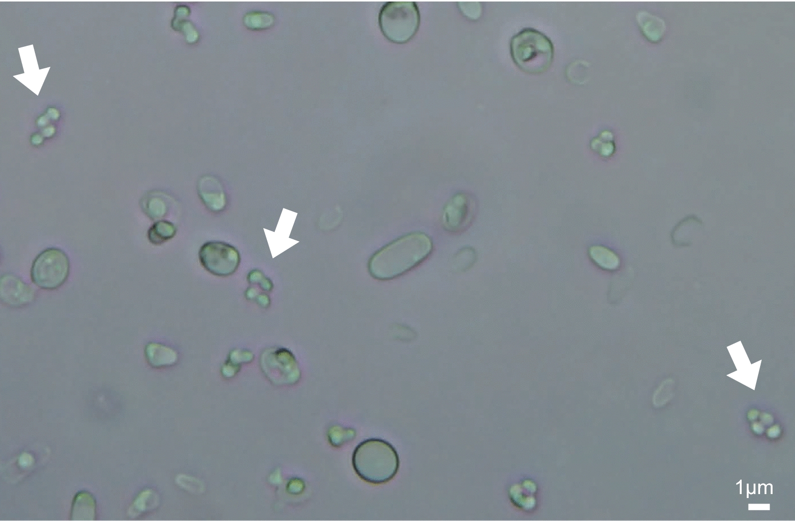
Bright field microscope image of a sporulated K. phaffii culture. Asci containing four spores are indicated with white arrows. Scale bar, 1 μm. This image is from a cross of CBS7435 × Pp2

Bright field microscope image of a sporulated K. phaffii culture. Asci containing four spores are indicated with white arrows. Scale bar, 1 μm. This image is from a cross of CBS7435 × Pp2