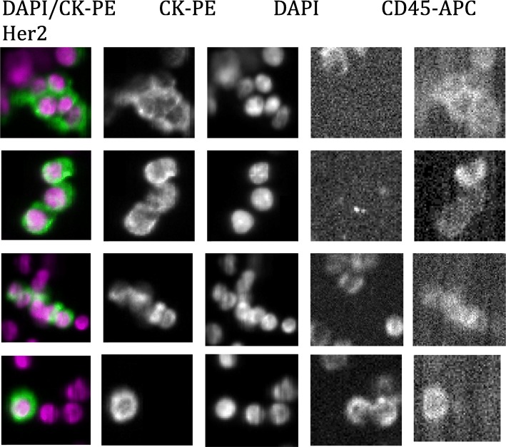
Fig. 4.
Representative images of a patient with CTC clusters. Shown are representative images of a patient with CTC clusters with nuclear (DAPI) and cytokeratin (CK-PE) staining. CD45 stains for non-CTC leukocytes. HER2/neu staining further distinguished CTCs from leukocytes in this patient. This sample was associated with the following ctDNA genomic alterations: CDH1, TP53, NF1, PIK3CB, BRCA1, CCND1, CDK6, FGFR1, MET, and MYC

