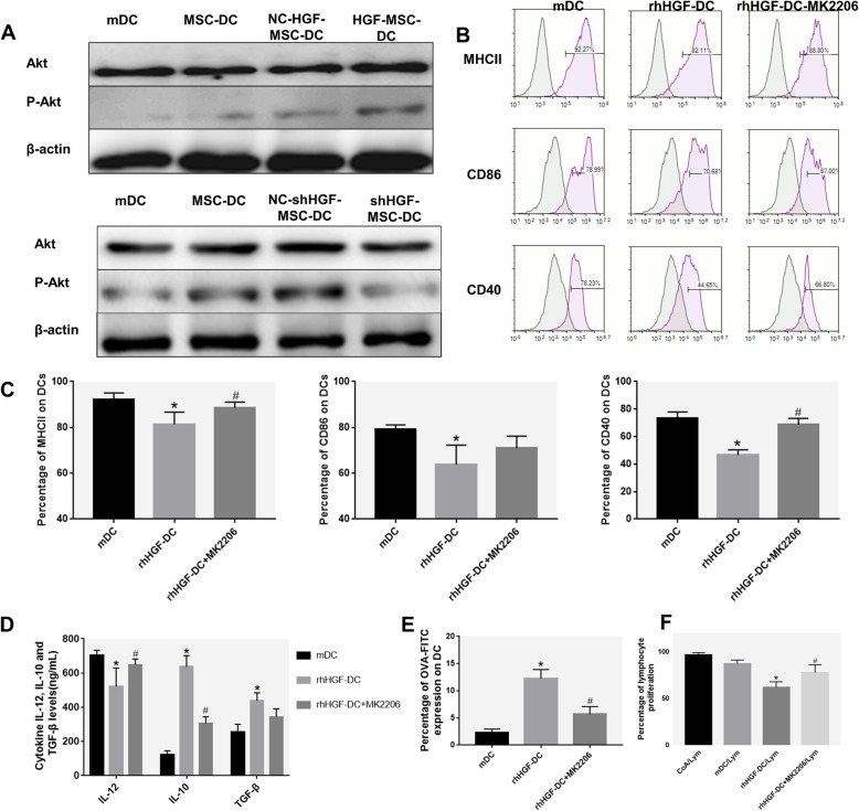
Fig. 5.
The role of the AKT pathway in MSC-secreted HGF induction of DC differentiation. a The expression of Akt protein and Akt phosphorylation levels in DCs cultured for 72 h in the presence of MSCs or MSCs after transduction was evaluated using Western blot analysis. b, c Immunophenotype analysis of DCs (expression of MHCII, CD86, and CD40 in mDCs cultured for 72 h in the presence of rhHGF or MK2206 and rhHGF). d Cytokine secretion profiles of DCs cultured for 72 h in the presence of rhHGF or MK2206 and rhHGF. e Phagocytic ability of HGF-induced DCs (percentage of OVA-FITC-positive cells). f Lymphocyte proliferation stimulated by ConA, mDCs, rhHGF-DCs, and rhHGF + MK2206-DCs. Normal BALB/c mouse solenocytes were used as responder cells in the mitogen proliferative assay. The proliferative responses were assessed by CFSE labeling and FACS (n = 3, *P < 0.05 versus mDC; #P < 0.05 versus rhHGF-DC; data are expressed as mean ± SD). Each experiment was repeated three times

