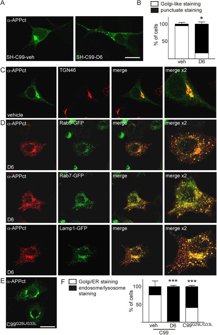
Fig. 4.
The subcellular localization of APP-CTFs is different in vehicle- and D6-treated C99-expressing SH-SY5Y and HeLa cells. a-b SH-SY5Y cells stably expressing C99 (SH-C99) were treated with or without D6 and immunostained with α-APPct. a APP-CTF immunostaining revealed a strong perinuclear staining typical of the Golgi in vehicle-treated cells and a punctuate staining in D6-treated cells. Bars in b correspond to the quantification of cells having either Golgi-like or punctuate APP-CTF immunostaining. A total of 400 cells for each condition from 3 independent experiments were counted. Data represent means ± SEM, *p < 0.05 as analyzed by the Mann Whitney U-test. c-f HeLa cells were transiently transfected with C99 and treated or not with D6 (c, d and f) or with C99G29L/G33L (e, f). c, Vehicle-treated C99-expressing cells presented a strong degree of colocalization of C99 (green) and the trans-Golgi apparatus marker TGN46 (red). d In D6-treated cells, the APP-CTF immunostaining (red) was punctiform and there was a high degree of colocalization with the subcellular markers Rab5-GFP, Rab7-GFP or Lamp1-GFP (green). Right panels show 2-fold high magnification images. e Cells transfected with C99G29L/G33L and APP-CTFs stained using α-APPct displayed a mix of Golgi-like and punctuate staining. f Bars correspond to the quantification of cells having either Golgi/ER or endosome/lysosome associated APP-CTF immunostaining. A total of 500 cells for each condition from 3 independent experiments were counted. Data represent means ± SEM, ***p < 0.001 as analyzed by 2-way ANOVA followed by Dunetts posthoc analysis

