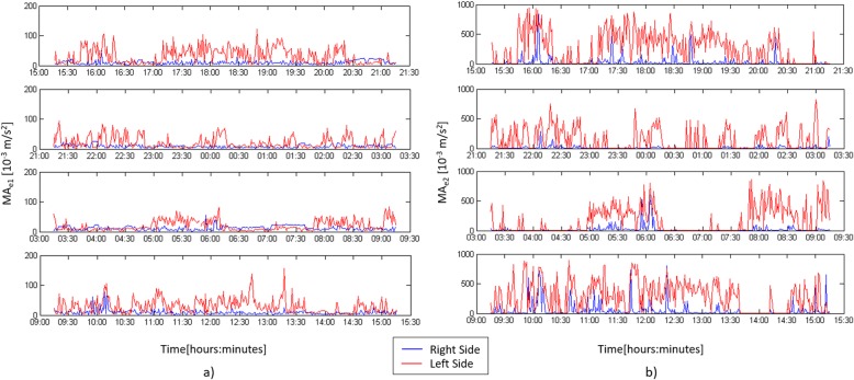
Fig. 2.
MAe1 (a) and MAe2 (b) profiles during the 24 h actigraphyc monitoring in a paradigmatic patient with right hemiplegic side: the blue line refers to the movement of the right wrist while the red profile to the left wrist. The figure illustrates the MAe1 and MAe2 profiles without excluding the recordings corresponding to passive movements due to nurses’ activities

