Fig. 7.

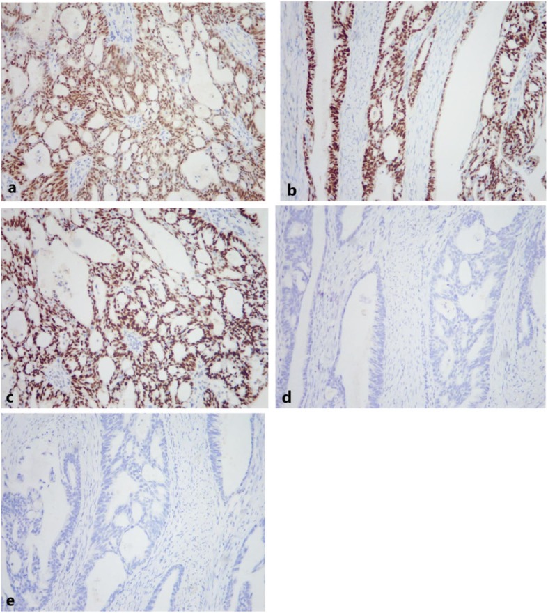
Immunohistochemical (IHC) staining of tumor cells for 5 markers (100×). a PAX-8 (positive). b ER (positive). c CK7 (positive). d CK20 (negative). e CDX-2 (negative)

Immunohistochemical (IHC) staining of tumor cells for 5 markers (100×). a PAX-8 (positive). b ER (positive). c CK7 (positive). d CK20 (negative). e CDX-2 (negative)