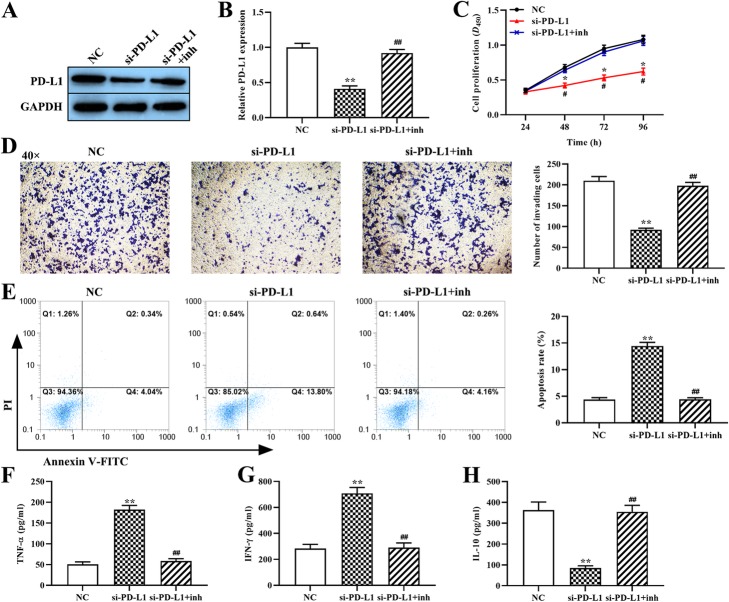
Fig. 4.
The effect of the miR-214–PD-L1 axis on the malignant behavior of OCI-Ly3 cells and on the cytokine secretion from T cells. (a and b) The expression of PD-L1 protein was determined using western blot. (c) The proliferation of OCI-Ly3 cells was determined using the CCK-8 assay. (d) The invasion ability of OCI-Ly3 cells was assessed using the Transwell assay (magnification, × 40). (e) The rate of OCI-Ly3 cell apoptosis was measured using flow cytometry. (f through h) The expression levels of TNF-α, IFN-γ and IL-10 were measured using ELISA. si-PD-L1: PD-L1 siRNA; si-PD-L1 + inh: PD-L1 siRNA + miR-214 inhibitor. *p < 0.05, **p < 0.01, compared with the NC group; #p < 0.05, ##p < 0.01, compared with the si-PD-L1 group

