Fig. 1.
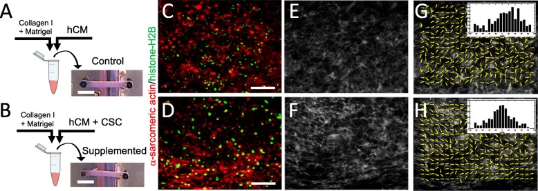
Fabrication of human engineered cardiac tissues (hECTs). a Diagram of fabrication of hECTs using a mix of hydrogel matrix (Collagen type-I plus Matrigel) and human induced pluripotent stem cell-derived cardiomyocytes (hCM). The solution is pipetted into a custom bioreactor with flexible end-posts, resulting in a self-assembled muscle strip-like shape within 72 h. b An identical approach was used to fabricate hCSC-supplemented hECTs, with the exception of adding cardiac stem cells (hCSCs) at a ratio of 90% hCM: 10% hCSCs. Scale bar of 2000 μm applies to a and b. c, d Two-photon images of control hECT (c) and supplemented hECT (d) stained for α-sarcomeric actin (red) and histone-H2B (green). Scale bar of 100 μm applies to panels c–h. e, f Second harmonic generation imaging of the corresponding two-photon image displaying collagen fibers, and g, h MatFiber3 analysis of collagen fiber orientation in images from e and f, with histograms of fiber alignment distribution (inset), showing mean angle ± CSD of 22.5° ± 33.5°, and − 2.95° ± 29.8°, respectively. Images were obtained with the hECTs oriented horizontally in the field of view

