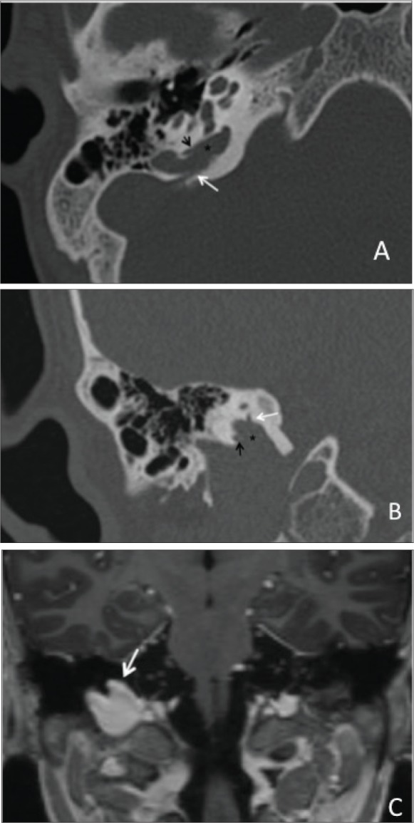
Figure 3.

Axial (A) and coronal (B) temporal bone CT in bone window demonstrates a superior lobulation (asterisk) emanating from the high ridIng jugular bulb. There are two focal bony dehiscences along the inferior limb of posterior SSC (black arrow) and vestibular aqueduct (white arrow). C: Coronal 3D T1 post contrast demonstrates a focal out-pouch of the right jugular bulb extending superiorly (arrow) consistent with a jugular bulb diverticulum.
