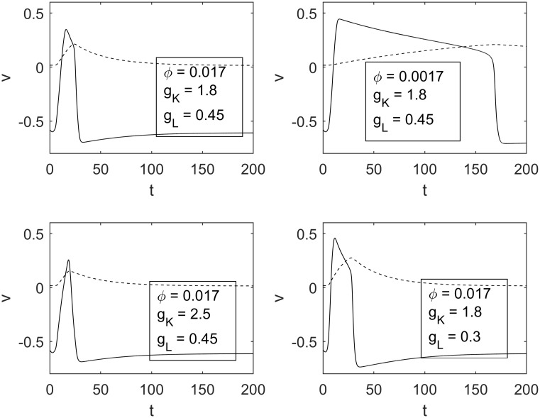
Fig 6. Various transmembrane potentials for different sets of parameters.
The top left panel depicts the transmembrane potential for the parameters in Table 1, while the top right, bottom left, and bottom right show transmembrane potentials for decreased ϕ, increased gK, and decreased gL, respectively.

