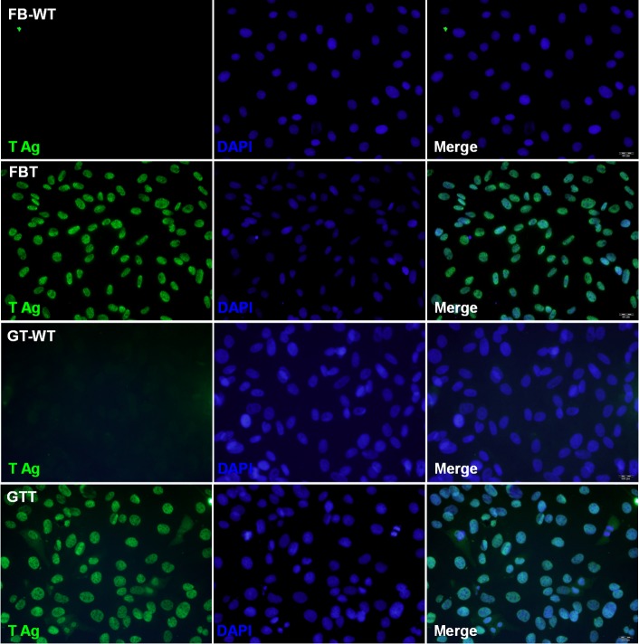
Fig 3. Immunofluorescence staining of goat fibroblast and testis cells.
Parental FB (FB-WT), GT (GT-WT), and the SV40 T transformed FBT, GTT cells were stained with the large T-specific antibody. Expression of SV40 T (labeled as T Ag) was shown as green fluorescence, and DAPI counterstaining indicates the cell nuclei.

