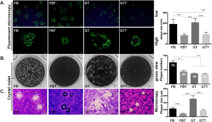
Fig 5. Plaque formation ability of ORFV in four different cell types.
The plaque formation ability was determined based on plaque assay. Cells were infected with OV20.0-GFP at 100 PFU/well and grown in an infectious medium containing 1.1% methylcellulose. Plaques were monitored either by fluorescence microscopy at both high and low magnifications (A), or stained with crystal violet at 6 dpi (B and C). The number of plaques was counted by the gross view (B, right panel), and the plaque size was measured under fluorescence microscopy (A, right panel) and bright field microscopy (C, right panel). The student T-test was performed to evaluate the plaque formation of the primary cells in comparison with its respective SV40-transformed cells against ORFV. A p-value < 0.05 was considered significant and is represented by the asterisk sign, e.g. *, **, *** indicated p-value < 0.05, 0.01, 0.001, respectively.

