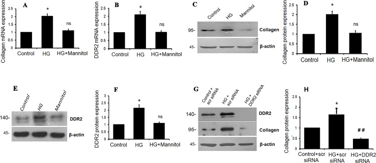
Fig 6. HG stimulates collagen type I and DDR2 in VSMCs.
(A-F) Sub-confluent cultures of VSMCs in M199 were stimulated with HG (25mM). (A) Collagen type I alpha 1 mRNA levels were determined by Taqman real-time PCR at 12 h post-HG treatment, with 18S rRNA as loading control. *p<0.05 vs. control, ns- not significant vs. control (One-way ANOVA p< 0.05). (B) DDR2 mRNA levels were determined at 12 h post-HG treatment. *p< 0.05 vs. control, ns- not significant vs. control (One-way ANOVA p< 0.05). (C and D) Collagen type I alpha 1 expression was determined by western blot analysis, with β-actin as loading control. *p<0.05 vs. control, ns- not significant vs. control (One-way ANOVA p< 0.05). (E and F) DDR2 protein expression was determined by western blot analysis. *p< 0.05 vs. control, ns- not significant vs. control (One-way ANOVA p< 0.05). (G and H) Regulatory relationship between DDR2 and collagen type I in HG-treated VSMCs. VSMCs were transiently transfected with DDR2 siRNA or scrambled siRNA. Following exposure of the transfected cells to HG for 12 h, collagen type I alpha 1 protein expression was examined by western blot analysis. DDR2 knockdown using siRNA was validated. *p< 0.05 vs control, ## p< 0.01 vs HG. (One-way ANOVA p< 0.05).

