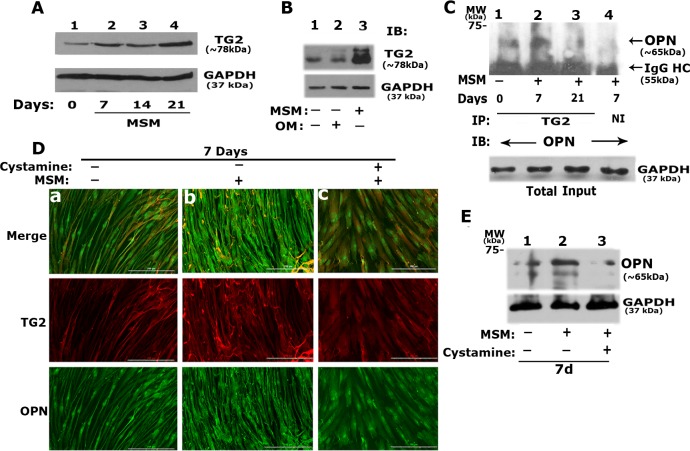
Fig 4. Effect of MSM on the interaction of TG2 with OPN in SHED cells treated with MSM in vitro.
A. Time-dependent effect of MSM treatment on the total protein level of TG2 was determined by immunoblotting (IB) analysis with an antibody to TG2. SHED cells were treated with MSM for 0, 7, 14, and 21 days (Fig a, lane 2–4). An equal amount of lysate protein (10 μg) was used. B. Comparison of TG2 levels in SHED cells incubated with OM (lane 2) or treated with MSM (lane 3) for 21 days. Cells grown in BM (lane 1) were used as controls. IB was conducted with an antibody to TG2. IB with an antibody to GAPDH was conducted after stripping in A and B, and used as a loading control. C. Analysis of the interaction of OPN with TG2 was determined by immunoprecipitation and IB analyses. An equal amount of protein lysates (10 μg) were used for immunoprecipitation with a TG2 antibody. Immunoprecipitates were subjected to IB with an antibody to OPN. IB of the total lysates with an antibody to GAPDH indicating the amount of protein from each sample were used for immunoprecipitation/IB analyses shown in panel C. D. Immunofluorescent analysis of the effect of TG2 inhibitor on the colocalization of TG2 and OPN in SHED cells treated with MSM (b) and MSM/Cystamine (c) for 7 days, Cells grown in BM (a) were used as controls. Imaging was conducted with the Cytation3 imager. Distribution of OPN and TG2 are shown either together (merge) or separately in green (OPN) and red (TG2) panels. Scale bar, 200μm. E. Immunoblotting analysis: The effect of cystamine on the cellular levels of OPN was determined in SHED cells treated with (lane 3) and without cystamine (lane 2) in the presence of MSM. Cells grown in BM (lane 1) were used as controls (-). GAPDH was used as a loading control. Data shown are representative of three independent experiments.

