. 2019 Nov 20;8:e50354. doi: 10.7554/eLife.50354

Figure 1. Cellular composition of the Larval brain by Single-Cell RNA-Seq.

(A) Experimental procedure. Drosophila late first instar larval brains/ventral nerve cords (VNC) were dissected and the VNCs were removed. Brains were collected and dissociated into a suspension of single cells. (B) Cell atlas of the larval brain reveals five main cell-types: neurons, NPCs, glial cells, UNs and other cell-types, represented in a Seurat UMAP plot. Groups are color coded. (C) Cell-types are recognized based on the expression of previously characterized marker-genes. A simplified heatmap illustrates this process: a subgroup of cholinergic neurons expresses the pan-neuronal marker elav and ChAT, a protein that catalyzes the biosynthesis of the neurotransmitter acetylcholine; NPCs are recognized by the expression of Notch and dpn and glial cells by repo expression. (D–D’’) Validation of the markers used to identify the different cell-types within the larval brain by immunostainings. The images display one lobe of the larval brain. Nuclei were labeled with DAPI and cellular borders with Dlg (disc large). Scale bar: 10 µm.

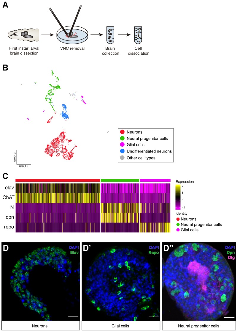
Figure 1—source data 1. Clusters properties.
Tables containing the top 10 differentially expressed genes across clusters, number of cells per cluster and the main literature in which the annotation processes were based on.

Figure 1.

Figure 1—figure supplement 1. Cell-type catalogue of the Larval brain.

Figure 1—figure supplement 1.

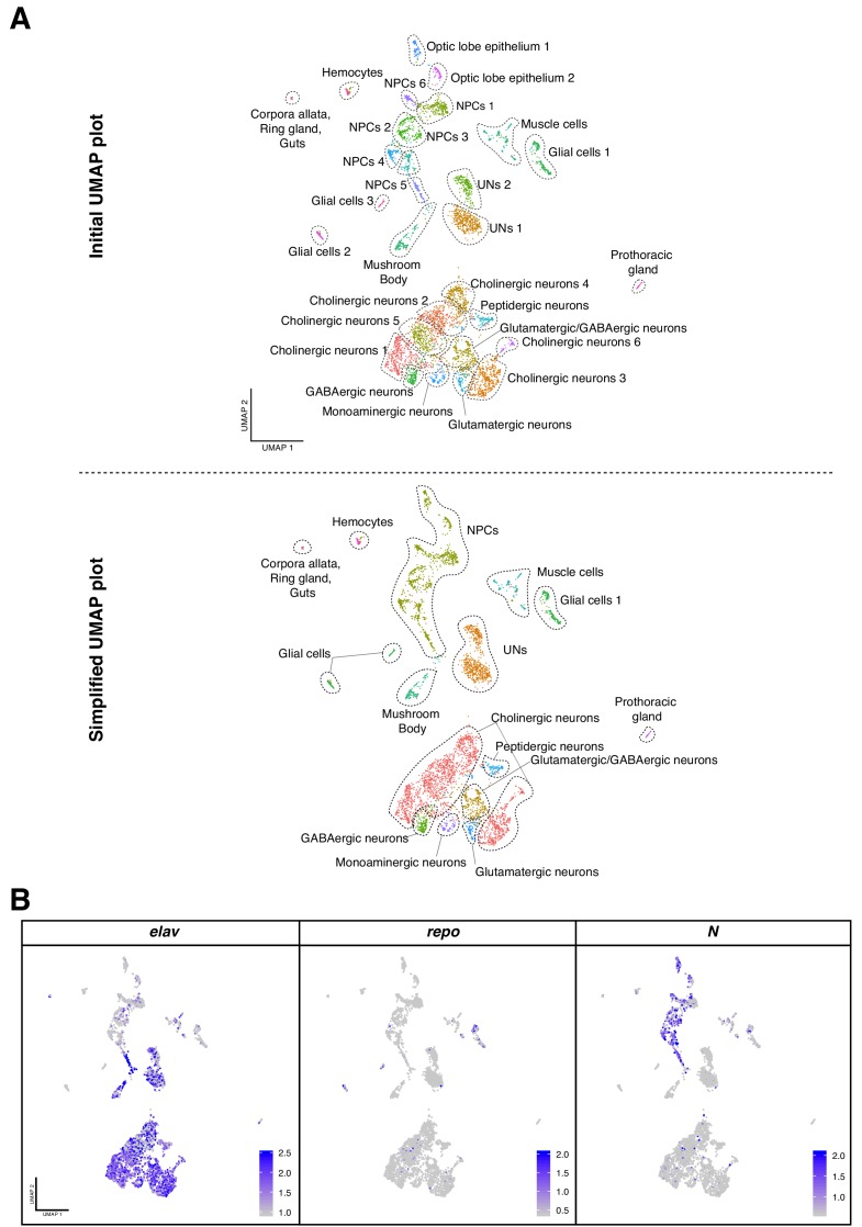
(A) Dimensional reduction, UMAP plot, of the total of cells considered for the analysis. Initially, 29 clusters were manually annotated (upper panel). Due to the similar expression profiles between different cell groups, clusters were combined to generate a simplified UMAP plot with 15 clusters of cells (lower panel). (B) The main cell-types identified are physically separated in the UMAP plot representation. Marker genes are used to confer cell identities: Elav for neurons, Notch for NPCs and repo for glial cells. Gene expression levels are color coded. NPCs: neural progenitor cells, UNs: undifferentiated neurons.
Figure 1—figure supplement 2. Non-neural cell-types identified in the dataset.

Figure 1—figure supplement 2.

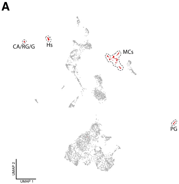
(A) Non-neural cells can be distinguished from the total number of cells. These cells are considered to be ‘contaminations’ and arise from reminiscent tissue attached to the brain during and after dissection: lymph gland, hemocytes, corpora allata, ring gland, gut and muscle (red cells). PG, prothoracic gland; CA/RG/G, corpora allata, ring gland and guts; Hs, hemocytes and MCs, muscular cells.