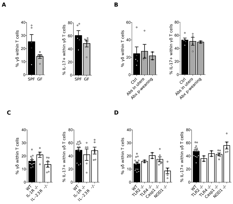
Figure 3. Meningeal γδ T cell homeostasis is independent of inflammatory signals.
Cell suspensions were prepared from the meninges of 8-12 weeks-old C57BL/6J WT mice, bred in a Specific Pathogen Free (SPF) (A-D) versus Germ Free (GF) (A) environment, treated or not with an antibiotic cocktail (B), and compared to IL-1R-/-, IL-23R-/- (C), TLR2-/-, TLR4-/-, Caspase 1-/- and NOD1-/- mice (D). Percentages of γδ T cells and IL-17 producers were analysed by FACS as illustrated in Fig. 1. Results are representative of 2-4 independent experiments.

