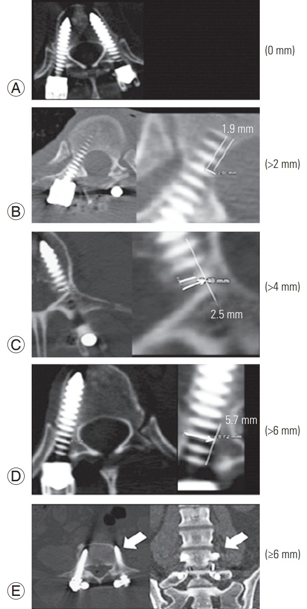
Fig. 1.

Computed tomography scans demonstrating the GertzbeinRobbins classification of screw positioning. The grades reflect the deviation of the screw from the ‘ideal’ intrapedicular trajectory. (A) Grade A is an intrapedicular screw without breach of the cortical layer of the pedicle; (B) grade B reflects a screw that breaches the cortical layer of the pedicle; however, it does not exceed it laterally by >2 mm. (C, D) Grades C and D indicate a penetration of <4 and <6 mm, respectively. (E) Grade E indicates a screw that does not pass through the pedicle or that, at any given point in its intended intrapedicular course, breaches the cortical layer of the pedicle in any direction by ≥6 mm (arrow). Reproduced from Schatlo et al. J Neurosurg Spine 2014;20:636-43, with permission from American Association of Neurological Surgeons [17].
