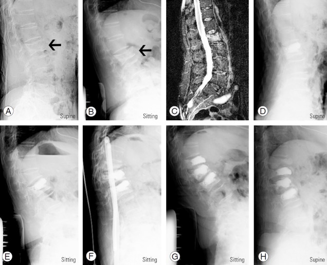
Fig. 3.
BKP-treated patient who developed an adjacent fracture. Lateral X-ray in supine (A) and sitting positions (B) and bone edema (C) on T2 fat-suppression magnetic resonance imaging when the patient visited our institution. The black arrow indicates a first lumbar vertebral fracture. (D) Lateral X-ray in supine position when she underwent BKP. Lateral X-ray in sitting position when she developed first (E), second (F), and third (G) adjacent vertebral fractures. (H) Lateral X-ray in supine position 1 year after the third adjacent vertebral fracture. BKP, balloon kyphoplasty.

