Figure 1.
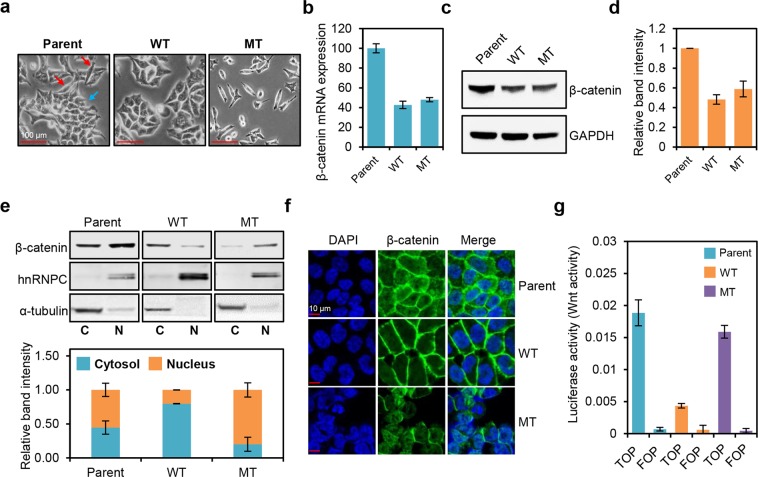
β-catenin mutation affects the localization and activation of β-catenin, as well as morphology of HCT116 cells. (a) Morphologies of HCT116 cells containing a wild-type (WT) β-catenin allele (HCT116-WT, WT), a mutant β-catenin allele (HCT116-MT, MT), or both wild-type and mutant alleles (HCT116-P, Parent). Red and blue arrows indicate mesenchymal-like and epithelial-like morphologies, respectively. (b) Relative β-catenin mRNA expression levels in HCT116 cell lines determined by qRT-PCR. (c) Western blot analysis of β-catenin protein levels in HCT116 cell lines. (d) Quantification of band intensities for the western blot shown in (c). (e) Top panel: western blot analysis of β-catenin levels in HCT116 cell lines subjected to cell fractionation. Bottom panel: quantification of band intensities for the western blot shown at top. hnRNPC and α-tubulin were used as nuclear and cytoplasmic markers, respectively. (f) Immunofluorescence microscopy analysis of β-catenin (stained in green) in HCT116 cells; DAPI staining is shown in blue. (g) Activation of WNT/β-catenin pathway in HCT116 cells, measured by TOP/FOP luciferase reporter assay. Error bars represent the standard deviation (SD) of the mean of two independent experiments. All assays shown in (c–f) were carried out in triplicate.

