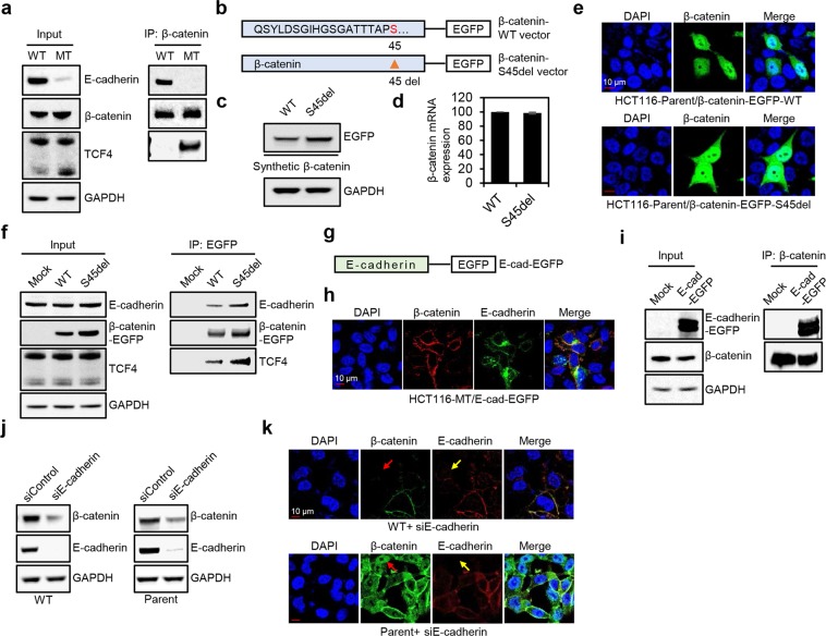
Figure 4.
E-cadherin binded to both WT and mutant β-catenin, and knockdown of E-cadherin downregulated membranous β-catenin expression. (a) Immunoprecipitation was performed in HCT116-WT and HCT116-MT cells using a β-catenin antibody. (b) Schematic model of EGFP-conjugated β-catenin expression constructs. β-catenin-S45del vector was generated by mutagenesis using β-catenin-WT vector. (c–e) Western blot, qPCR, and immunofluorescence microscopy analysis of HCT116-P cells transfected with β-catenin-S45del vector or β-catenin-WT vector. (f) Immunoprecipitation was performed using an EGFP antibody in HCT116-P cells transfected with a control vector or β-catenin expression vectors. (g) Schematic model of EGFP-conjugated E-cadherin expression vector. (h,i) Immunofluorescence microscopy analysis and immunoprecipitation with a β-catenin antibody was performed in HCT116-MT cells transfected with E-cadherin expression vector. (j,k) Western blot and immunofluorescence microscopy analysis of HCT116-WT and HCT116-P cells transfected with siRNA against E-cadherin. Red and yellow arrows indicate loss of WT β-catenin and loss of E-cadherin, respectively. All assays were carried out in triplicate.

