Figure 7.
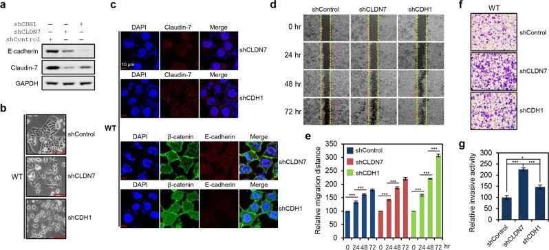
Loss of tight junctions mediated by Claudin-7 dysregulation was sufficient for acquisition of mesenchymal-like features by HCT116 cells. (a) Western blot analysis of E-cadherin and Claudin-7 expression in HCT116-WT cells with stable shRNA-mediated knockdown of Claudin-7 (shCLDN7) or E-cadherin (shCDH1). (b) Morphological changes in HCT116-WT cells after stable knockdown of Claudin-7 or E-cadherin. Cell images were obtained using the 20x objective. (c) Immunofluorescence microscopy analysis of Claudin-7 (stained in red), β-catenin (stained in green), and E-cadherin (stained in red) in HCT116-WT cells with stable shRNA-mediated knockdown of Claudin-7 or E-cadherin knockdown. (d) Wound-healing assay performed using HCT116-WT cells with stable knockdown of Claudin-7 or E-cadherin. Gap closure was measured at 24, 48, and 72 h after the initial scratch, due to the relatively low migratory activity of HCT116-WT. (e) Quantification of relative migration distances for the wound-healing assay shown in. (d) Statistical significance between 0 and 24 hours, 24 and 48 hours, and 48 and 72 hours is shown. (f) Invasion assay performed using HCT116-WT cells with stable knockdown of Claudin-7 or E-cadherin. Invasive activity was measured after 48 h by crystal violet staining. (g) Quantification of the relative number of stained knockdown cells in (f) using ImageJ software. Statistical significance between shControl, shCLDN7, and shCDH1 is shown. Error bars in (e,g) represent the SD of the mean of results from three independent experiments. All assays were carried out in triplicate.

