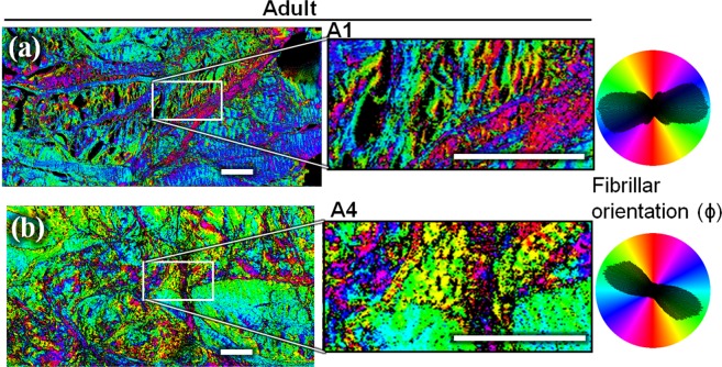
Figure 4.
Collagen fibrils orientation in the imaging plane (ϕ) measured by P-SHG, for the same two adults’ specimen (A1 (a) and A4 (b)) as in Fig. 2. These mature menisci have an organized structure, with some thick fibers that clearly delimitate bundles of homogeneous fibrils. On the polar histograms, the orientation shows a principal component coupled with some other components corresponding to the numerous – but still limited – orientations. Scale-bars: 200 μm.

