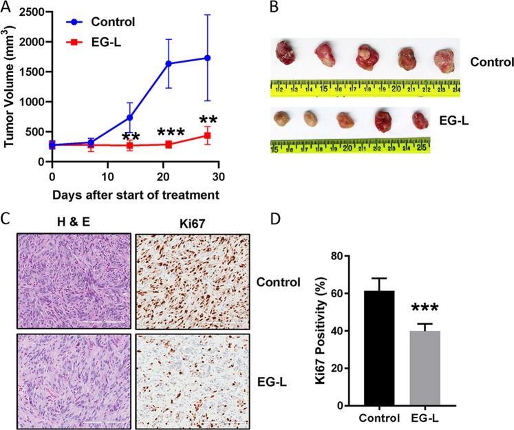
Fig. 4.
Validation of the result obtained from single mouse trial in cohorts of five mice. a 5 × 10⁶ 786-O cells were subcutaneously injected into the right flanks of 8-week-old male SCID mice. Tumors were allowed to grow until the average tumor size is ~300 mm3. Then mice were treated with vehicle or EG-L (n = 5 mice per treatment group) 3× per week for 4 weeks. Tumors were measured weekly and tumor volume was plotted to obtain the respective growth curves. EG-L demonstrated significant inhibition compared to the vehicle group. ** and *** denote p < 0.01 and p < 0.001 compared to control, respectively. b Images of the harvested tumors at the end of the experiment. c Representative images of H&E and Ki67 staining of the tumor tissue sections. Bar length = 200 µm. d Quantification of Ki67-positive nuclei. *** denotes p < 0.001 compared to control (n = 3 tumors per group, five spatially different regions from each tumor section).

