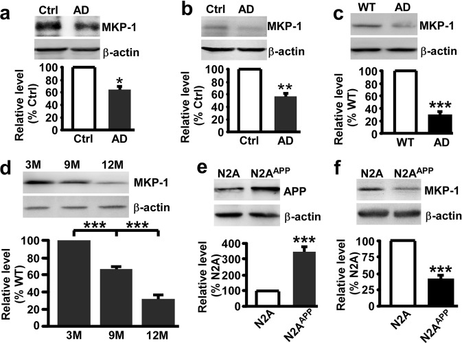
Fig. 1.
MKP-1 was decreased in AD. a, b The protein level of MKP-1 assessed by western blot in the hippocampus a and temporal cortex b of control (Ctrl) and AD patients. *p < 0.05 by unpaired Student’s t test. n = 4–6 in each group. c The protein level of MKP-1 in the hippocampus of wild-type (WT) and APP/PS1 transgenic AD model mice at the age of 9 months. ***p < 0.001 by unpaired Student’s t test. n = 6 in each group. d The protein level of MKP-1 in the hippocampus of AD mice at different ages. ***p < 0.001 by one-way ANOVA. n = 3 in each group. e, f The protein levels of APP e and MKP-1 f assessed by western blot in lysates of N2A and N2AAPP cells. ***p < 0.001 by unpaired Student’s t test. n = 3–6 in each group.

