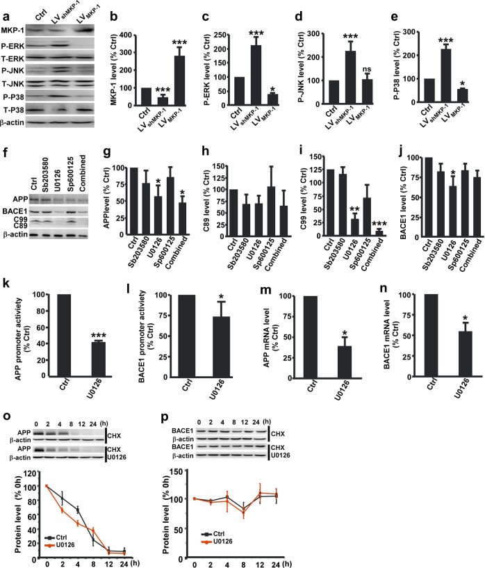
Fig. 4.
MKP-1 inhibited APP processing through inactivation of the ERK/MAPK signaling pathway. a–e Immunoblot of the expression of MKP-1 b, P-ERK c, P-JNK d and P-P38 e in lysates of N2AAPP cells after overexpression of MKP-1 by LVMKP-1 or knockdown of MKP-1 by LVshMKP-1. *p < 0.05 and ***p < 0.001 by one-way ANOVA. n = 4–5 in each group. f–j Immunoblot of the expression of APP g, C89 h, C99 i and BACE1 j in lysates of N2AAPP cells treated with the ERK inhibitor U0126, the JNK inhibitor SP600125, the P38 inhibitor SB203580 or the three inhibitors together (Combined). *p < 0.05, **p < 0.01 and ***p < 0.001 by one-way ANOVA. n = 5–7 in each group. k, l The mRNA levels of human APP k and BACE1 l assessed by qPCR in N2AAPP cells and N2AAPP cells treated with U0126. ***p < 0.001 for APP and *p = 0.032 for BACE1 by unpaired Student’s t test. n = 3–4 in each group. m, n The promoter activity of APP m and BACE1 n assessed by luciferase assay in N2AAPP cells and N2AAPP cells treated with U0126. *p = 0.036 for APP and *p = 0.040 for BACE1 by unpaired Student’s t test. n = 3–4 in each group. o, p The degradation of APP o and BACE1 p as assessed by half-life measurements in the N2AAPP cells after U0126 treatment. p = 0.264 for APP and p = 0.847 for BACE1 by two-way ANOVA. n = 4–5 in each group.

