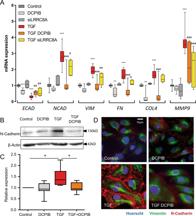
Fig. 5. DCPIB or LRRC8A knockdown alters the expression of EMT markers after TGFβ1 treatment in HK-2 cells.
a Fold increase in mRNA levels of ECAD (E-cadherin), NCAD (N-cadherin), VIM (Vimentin), FN (Fibronectin), COL4 (Collagen IV) and MMP9 (Matrix Metalloproteinase-9) in HK-2 cells cultured with or without TGFβ1 (2.5 ng/ml) for 24 h in the presence or absence of DCPIB (20 µM) or after silencing of LRRC8A (siRNA). 36B4-normalized mRNA levels in control cells were used to set the baseline value at unity. Box plots illustrating the mRNA fold increase of 5–13 experiments from five independent cell cultures. Kruskal-Wallis with Dunn’s multiple comparison post hoc test was used with **p < 0.01, ***p < 0.001 vs control; #p < 0.05, ##p < 0.01, ###p < 0.001 vs TGF. b, c Protein expression of N-cadherin in cells treated with TGFβ1 (2.5 ng/ml) for 24 h in the presence or absence of DCPIB (20 µM). β-actin was used as a loading control. Representative Western blots (b) and corresponding quantitative analysis (c) performed on five independent experiments. The results are expressed as the n-fold increase over the control and Friedman + Dunn statistic test was used with *p < 0.05. d Immunofluorescence staining of N-cadherin and vimentin proteins. Nuclei were stained with Hoechst 33342 dye. Cells were treated with or without TGFβ1 (2.5 ng/ml) for 24 h in the presence or absence of DCPIB (20 µM) as indicated. Scale bar: 10 µm.

