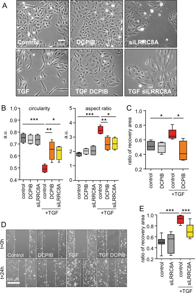
Fig. 6. Pharmacological inhibition of VRAC or LRRC8A knockdown attenuates TGF β1-induced cell morphology changes, migration and invasion.
a Representative micrograph (phase-contrast microscopy) illustrating the morphological changes induced by TGFβ1 treatment (2.5 ng/ml, 24 h) and the inhibitory effects of DCPIB (20 µM) or LRRC8A gene silencing. b Box plots illustrating variations in circularity (left) and aspect ratio (right) as two indicators of morphological changes induced by TGFβ1 treatment concomitant with DCPIB exposure or LRRC8A knockdown (siRNA). The two parameters were analyzed with ImageJ shape descriptor software. Data were obtained from 60 cells (four independent experiments). ANOVA with Bonferroni’s multiple comparison post hoc test statistical test was used with *p < 0.05, **p < 0.01. c, d Box plots (c) and micrographs (d) illustrating the effect of pharmacological inhibition of LRRC8/VRAC in migration processes using wound healing experiments. HK2 cells were scratched and wounded after a 24 h starving period without serum. Wound closure was monitored using an inverted microscope for 24 h with or without TGFβ1 (2.5 ng/ml) and DCPIB (20 µM). Scale bar: 500 µm. Wound closure was quantified using ImageJ software 24 h after wounding. Values are expressed as the ratio of the initial wound area and were obtained from six measurements (three independent experiments). Kruskal-Wallis with Dunn’s multiple comparison post hoc test was used with *p < 0.05. e Independent series of wound healing assay were performed in HK2 cells to illustrate the effect of LRRC8A silencing (siLRRC8A) in migration processes (six measurements from two independent experiments; Kruskal-Wallis with Dunn’s multiple comparison post hoc test with ***p < 0.001).

