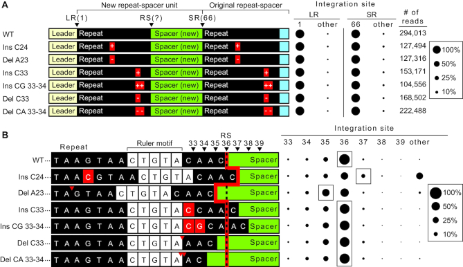
Figure 8.
Ruler-based mechanism influences second-site integration in vivo. (A) (left panel) Annotated sequence of expanded CRISPR array in vivo with single and double insertion and deletion mutations in the repeat sequence. (Right panel) Sites of integration represented as percent total of mapped reads at the leader-repeat junction (LR) and spacer–repeat junction (SR). (B) (left panel) Sequences spanning the repeat–spacer junction and site of integration. Red line highlights expected site of integration relative to insertions and deletions upstream and downstream of the ruler element (5′-CTGTA-3′) as indicated by red arrows or boxes. (Right panel) Integration site at the positions spanning the repeat–spacer junction.

