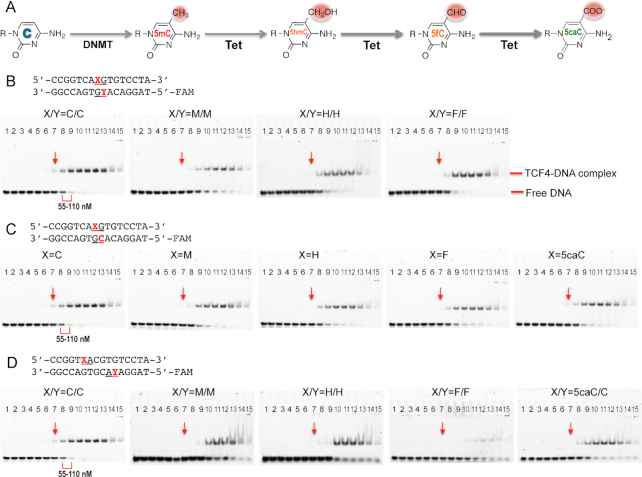
Figure 2.
Electrophoretic mobility shift assay of TCF4 bHLH protein binding to oligos containing a single E-box. (A) Schematic of chemical reactions of DNA cytosine methylation by DNMT and 5mC oxidations by Tet enzymes. (B) The central CpG dinucleotides are unmodified (C/C) or fully modified (M/M, H/H, F/F; where M = 5mC, H = 5hmC, and F = 5fC). (C) The central CpG dinucleotides are hemi-modified (M/C, H/C, F/C or 5caC/C; where 5caC = 5-carboxyC). (D) The two outer CpA dinucleotides are unmodified (C/C), fully modified (M/M, H/H, F/F) or hemi-modified (5caC/C). The protein concentrations used were a maximum of 7 μM (the right most lane 15 of each panel) followed by serial 2-fold dilutions (from right to left). The arrows indicated a reference point where the shift was observed for the unmodified oligo. The same samples were quantified by fluorescence polarization (Supplementary Figure S1).

