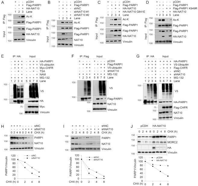
Figure 7.
NAT10 acetylates PARP1 at K949 and enhances its stability. (A–D) HEK293T cells were transfected with the indicated expression vectors. After 48 h of transfection, lysates were subjected to IP and immunoblotting analyses with the indicated antibodies. The immunoprecipitated PARP1 has been adjusted to be equal to make the levels of acetylated PARP1 comparable to those of immunoprecipitated PARP1. (E–G) HEK293T cells were transfected with the indicated expression vectors. After 48 h of transfection, cells were treated with 10 μM MG-132 for 6 h and lysates were subjected to IP and immunoblotting analyses with the indicated antibodies. Lysis buffer was also supplemented with 10 μM MG-132 in the subsequent assays. (H) MCF-7 cells were transfected with siNC or siNAT10. After 48 h of transfection, cells were treated with 100 μg/ml CHX for the indicated times and analyzed by immunoblotting. Relative PARP1 levels (PARP1/Vinculin) are shown in lower panel. (I) MCF-7 cells stably expressing shNC or shNAT10 were treated with 100 μg/ml CHX for the indicated times and analyzed by immunoblotting. Relative PARP1 levels (PARP1/Vinculin) are shown in lower panel. (J) HEK293T cells were transfected with pCDH or HA-NAT10. After 48 h of transfection, cells were treated with 100 μg/ml CHX for the indicated times and analyzed by immunoblotting. Relative PARP1 levels (PARP1/Vinculin) are shown in lower panel. In panels (H) to (J), quantitative results are represented as mean ± s.d. as indicated from three independent experiments.

