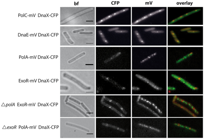
Figure 2.

Altered localization patterns after induction of DNA damage. Localization by epifluorescence in representative live Bacillus subtilis cells after induction of DNA damage with MMC. The overlay panels show DnaX-CFP in red and PolC-mV, DnaE-mV, PolA-mV, and ExoR-mV in green. Note that DnaX-CFP foci rarely colocalize with PolA-mV or ExoR-mV foci. Scale bar: 2 μm.
