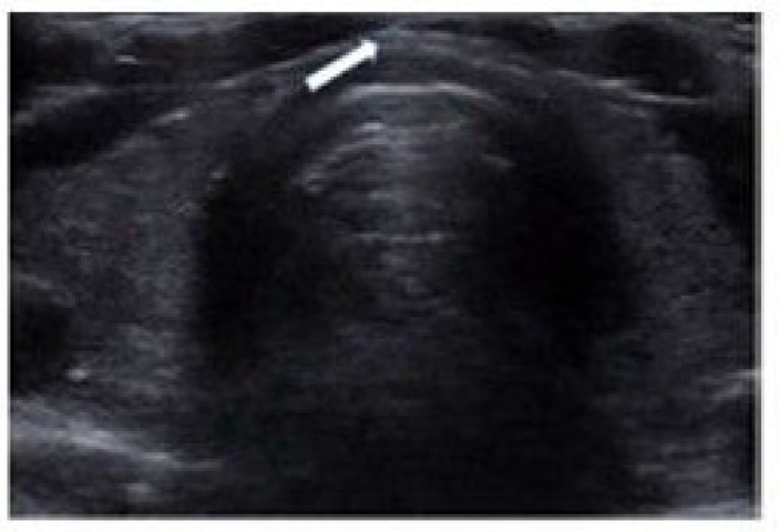
Venous Doppler ultrasonography images of lower extremity demostrating May-Thirner syndrome anatomy with compression of the left iliac vein via the right common iliac artery

Venous Doppler ultrasonography images of lower extremity demostrating May-Thirner syndrome anatomy with compression of the left iliac vein via the right common iliac artery