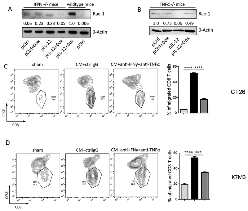
Fig. 4. Inflammatory cytokines IFNγ and TNFα facilitate CD8+ T cell migration to tumor cells.
(A, B) LLC tumor–bearing IFNγ−/− or TNFα−/− mice (n=3/group) were treated twice (10 days apart) with one of the four indicated treatments: control, doxorubicin (Dox), IL-12–encoding DNA, or IL-12–encoding DNA plus doxorubicin. Tumors were collected on day 4 after the second treatment. Tumors were subjected to immunoblotting of Rae-1 in comparison with wildtype littermates. Values under each band indicate the density of protein expression. (C, D) CT26 (C), K7M3 (D) tumor cells were cultured for 24 h in regular RPMI medium or with T cell–conditioned medium (CM) with or without anti-IFNγ (1 μg/mL) and anti-TNFα (0.5 μg/mL) neutralizing antibodies. CFSE-labeled tumor cells were plated in the lower chambers of Transwell plates, while activated CD8+ T cells (5 × 10⁵/well) were plated in the upper wells of the insert. After 24 h of co-culture, CD8+ T cells that had migrated to the lower chambers were detected via flow cytometry. Results are representative of three repeated experiments. ** P<0.01; *** P<0.005; **** P<0.001.

