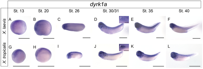
Figure 2. In situ hybridization of dyrk1a across developmental stages demonstrates kidney expression in X. laevis and X. tropicalis.
To demonstrate spatial-temporal expression of dyrk1a in the kidney, in situ hybridization was performed. Given that the RNA probe was designed against the X. tropicalis sequence, both species were analyzed. Pronephric kidney development occurs between stages 12.5-40. Expression of dyrk1a can be visualized in stage 31-40 embryos suggesting Dyrk1a may be important for kidney development. For clarity, insets (D’ and J’) for stage 30/31 tadpole kidneys with 200μm scale bars have been added. All other scale bars represent 1000μm.

