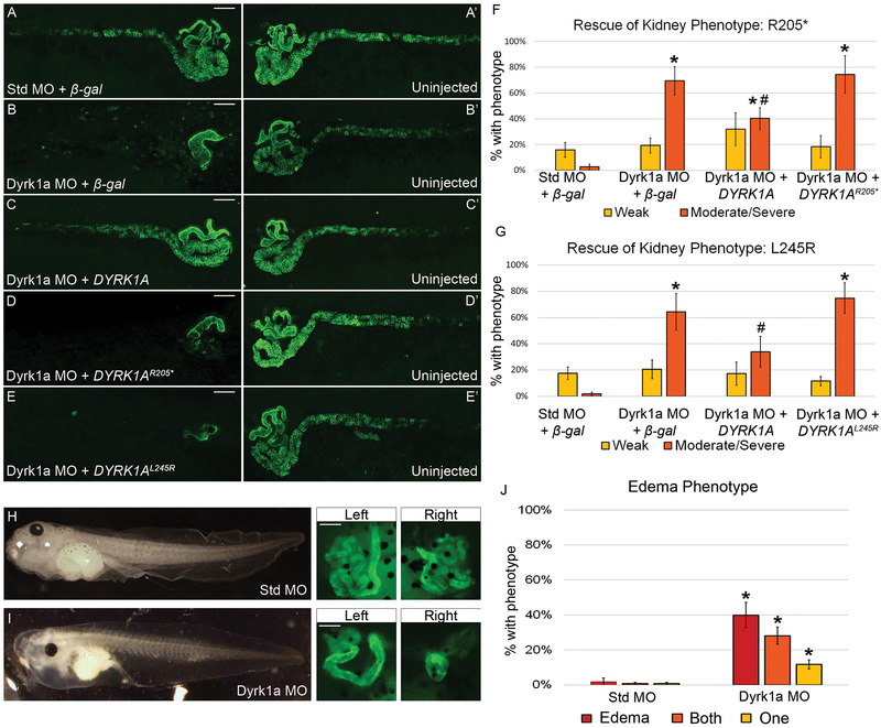
Figure 3. Loss of Dyrk1a affects kidney development in Xenopus laevis.
(A-E’) Embryos were unilaterally injected at the 8-cell stage with 10ng of Dyrk1a MO or Standard MO (Std MO) along with 50 pg β-gal, wild-type, DYRK1AR205*, or DYRK1AL245R RNA. Stage 40 tadpoles were stained with kidney antibodies 3G8, which label the proximal tubules and 4A6, which labels the distal and connecting tubules. Letters without apostrophes (A-E) represent the injected side, whereas letters with apostrophes (A’-E’) represent the uninjected side. (B) Knockdown with a translation-blocking Dyrk1a MO disrupts kidney development which can be partially rescued (C) by co-injecting with wild-type human DYRK1A RNA but not (D-E) DYRK1AR205* or DYRK1AL245R RNA. (A) Co-injection of a Standard MO and β-gal serve as a negative control. Scale bars represent 100 μm. (F) The graph demonstrates a significant difference between embryos injected with either Dyrk1a MO + β-gal or Dyrk1a MO + DYRK1AR205* versus with Dyrk1a MO + DYRK1A suggesting successful rescue with human DYRK1A but not with the nonsense RNA. (G) The second graph demonstrates a significant difference between embryos injected with Dyrk1a MO + DYRK1AL245R versus with Dyrk1a MO + DYRK1A, which suggests that the missense RNA also fails to rescue. Significance was established against embryos that had a moderate or severe kidney phenotype (orange bar) and excluded embryos that had a weak phenotype (yellow bar). (F) * (asterisk) = p<0.001 comparing individual experimental groups to Standard MO + β-gal. # (pound sign) = p<0.006 comparing Dyrk1a MO + DYRK1A to Dyrk1a MO + DYRK1AR205*. (G) * (asterisk) = p<0.001 comparing Standard MO + β-gal to Dyrk1a MO + β-gal or Dyrk1a MO + DYRK1AL245R. # (pound sign) = p<0.05 comparing Dyrk1a MO + DYRK1A to Dyrk1a MO + DYRK1AL245R. For edema assays, embryos were injected at the 4-cell stage in both ventral cells to target both kidneys while avoiding the dorsal cells fated to become the heart and liver, which can also lead to edema. (H) Embryos injected with the Standard MO did not develop edema while embryos injected (I) with the Dyrk1a MO did develop edema and also suffered from abnormal kidney formation. (J) The graph demonstrates a significant difference in edema and kidney abnormalities in embryos injected with either Standard MO or Dyrk1a MO. * (asterisk) = p<0.008 comparing Standard MO to Dyrk1a MO embryos with edema, defects in one, and defects in both kidneys. Error bars represent standard error. For ease of comparison of (n) and p-values across conditions, please refer to Table S1-3.

