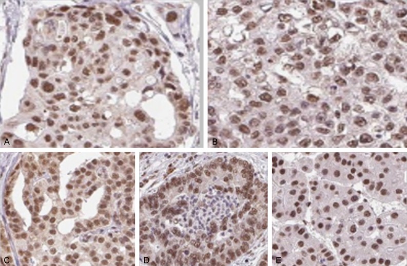
Figure 2.

Antibody staining of five standard cancer tissues samples highlighting the localization of SNRPG in tumor cells. A. Colorectal Cancer. B. Breast Cancer. C. Prostate Cancer. D. Lung Cancer. E. Liver Cancer. Antibodies are labeled with DAB (3,3’-diaminobenzidine) and the resulting brown staining indicates where an antibody has bound to its corresponding antigen (SNRPG). Staining: Medium, Intensity: Moderate, Quantity: > 75%, Location: Nuclear, Magnification: 40 × (Figure taken from [18]).
