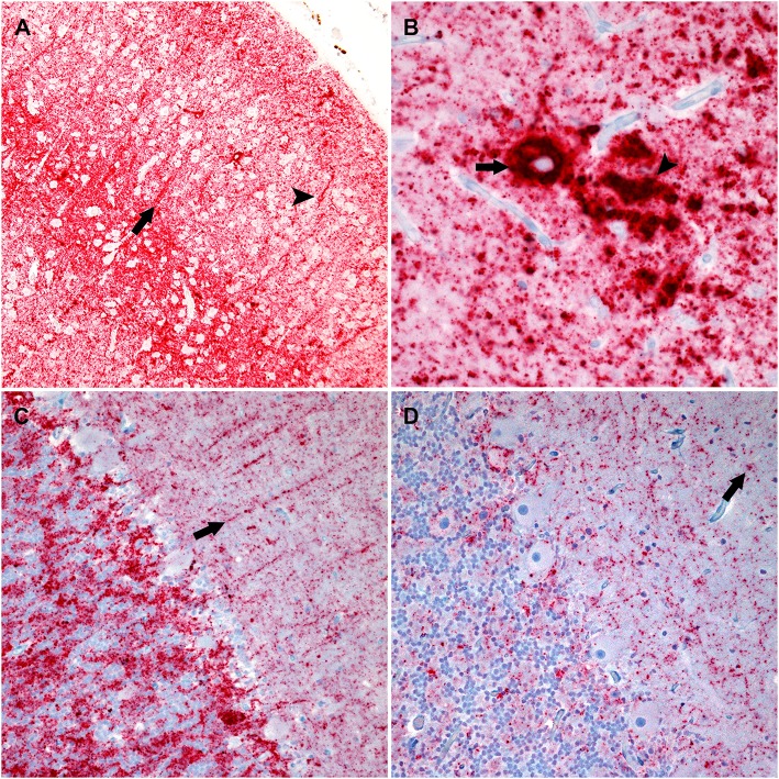
Figure 2.
Photomicrographs of brain regions from sheep that were inoculated intracranially with the agent of bTME. Immunohistochemistry for PrPSc, monoclonal antibodies F89/160.1.5, and F99/97.6.1. (A) PrPSc in the frontal neocortex from a sheep with an ARQ/ARR genotype. In this genotype there is diffuse particulate that is denser in neocortical layers IV and V. There also are prominent linear (arrow) and perineuronal (arrowhead) immunolabeling patterns. (B) In the cerebrum from sheep with the ARQ/ARR genotype, there are vascular plaque-like accumulations (arrow) of PrPSc. The arrowhead indicates an area of coalescing particulate immunolabeling. (C) Cerebellum from a sheep with an VRQ/ARQ genotype. In the granular layer there is particulate and intraglial immunolabeling, and in this image, linear type PrPSc (arrow) is evident in the molecular layer. The immunolabeling types present are typical of the VRQ/VRQ, VRQ/ARQ, and ARQ/ARQ genotypes. (D) Cerebellum from a sheep with the ARQ/ARR genotype. Overall, there is less PrPSc immunolabeling. In the selected image, there is primarily diffuse particulate with intermittent intraglial (arrow) PrPSc.

